
Dr. Kevin Park continues to look into different parts of the the central nervous system (CNS) to shed light on how the obstinate signal conducting neurons encased in our skeleton differ from those in the peripheral nervous system (PNS) nerves that project outside of our skulls and spines. Dr. Park’s research program aims to learn about CNS regeneration by studying optic nerve (“retinal ganglion cells”) injury and regeneration, and his efforts are endorsed by two new National Institute of Health (NIH) grants.
The link between optic and spinal nerves might seem illusive at first, but start by considering the almost inseparable bond between vision and movement. No plants have eyes, and within animals it is a general rule that acuity of vision is proportional to speed of movement—take for instance the Peregrine falcon’s >200 mi/hr top speed and legendary vision, or the Mantis shrimp’s “world’s fastest punch” and immensely complex eyes to match. Furthermore, despite difference in our perceived experience of vision and movement there are certain surprising similarities between the kinds of nerves that originate from within the skull and spine. First, these CNS nerves are infamously resistant to regeneration, one of the reasons why CNS trauma leading to blindness and paralysis are often, at least for the time being, chronic conditions in adults. Beyond plasticity, these nerves operate best in “small-world networks” characterized by select long-distance connections that enhance efficiency at the risk of having vastly separated the signal from its target should the connection be lost. Furthermore, the similarities between CNS neurons extend beyond the signal conducting cells themselves. All nerves, both CNS and PNS, are embedded in a matrix of support cells known as glia. The glial cells that ensheath—or “myelinate”—optic and spinal nerves are called oligodendrocytes, while those that wrap PNS nerves are called Schwann cells. Fundamental differences between oligodendrocytes, that wrap multiple cell axons, and Schwann cells, that prefer to keep their ensheathing to one nerve at a time, is another shared characteristic between optic and spinal nerves that might make it so that breakthrough regenerative discoveries from one of these cell-types might guide regenerative therapies in many CNS nerves.
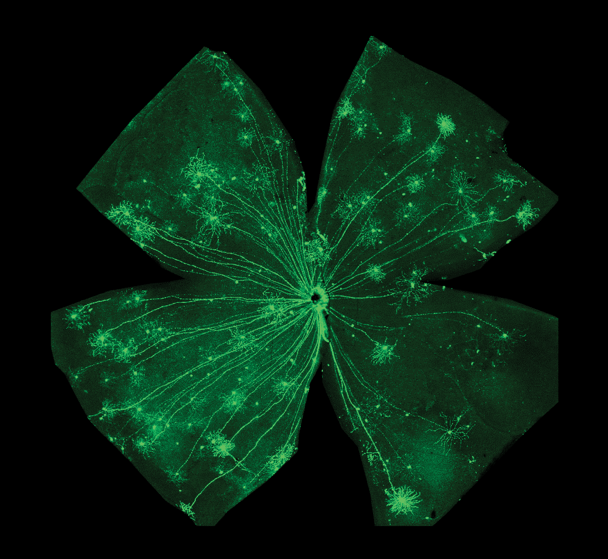
It is on this premise Dr. Park’s new NIH grants function, with their findings directly applicable to visual disease and injury but also with his optic nerves decidedly acting as a model for studying regeneration of long-distance axonal connections in the CNS writ large. Dr. Park’s first grant, an exploratory grant intended to fund high-risk/high-reward projects in the conceptual phase of development, aims to develop a rodent model of glaucoma whereby optic nerve axons are damaged by elevated intraocular pressure instead of by surgical damage of the nerve. This approach mirrors that of the “contusion vs transection” model of experimentally inducing spinal cord injury in animals, with contusion reflecting the real-world neurotrauma causing CNS paralysis in people. The second, and larger, grant is an ambitious application of a suite of cutting edge technologies to Dr. Park’s tried-and-true optic nerve model to identify regeneration-promoting factors that can be the target for novel treatment. This project combines a whole-brain imaging technique and single-cell tracing so that individual, intact optic nerve cells can be visualized in three dimensional space from origin to target. It has previously been shown that certain factors can stimulate optic nerve regeneration, but due to the long-distance projections it has not yet been possible to connect the exact signal generating nerves on one side of the injury to the targets they grow to on the regenerated side. Dr. Park’s approach will allow for one-to-one 3D identification of the regenerating optic nerves, and once these nerves are identified they can be studied to determine the type of molecular properties that drive their regenerative behavior.