
Miami Project scientists, Pantelis Tsoulfas, M.D., Associate Professor at the Department of Neurological Surgery and The Miami Project to Cure Paralysis, and colleagues in collaboration with the Murray Blackmore laboratory at Marquette University, recently presented a paper in bioRxiv (pronounced “bio-archive”) on Brain-wide Quantification of the Supraspinal Connectome. The researchers, while they await peer-review publication, wanted to share this important work, primarily done during the pandemic, because it shares a comprehensive and accessible approach to obtain detailed information about the number and location of descending projection neurons throughout the mouse brain. This information provides an effective tool to disseminate a detailed understanding of the supraspinal (above the spinal cord and vertebral column, most commonly talking about the brain) connectome (the structural connectivity of the nervous system), and a needed platform to rapidly achieve brain-wide profiling of disruption and restoration of brain and spinal cord connectivity after injury. As a result of this work, the researchers created a website dedicated to the data so scientists anywhere can explore, visualize and quantify the supra spinal neurons with projections to the cervical, thoracic and lumbar segments. The new site is called www.3dmousebrain.com/ is free and open to all users.
“The main reason for starting this project is that during our studies we knew that we are neglecting the majority of neurons that project to the spinal cord because, the experimental tools used so far have certain limitations. It is important to understand how our therapeutic interventions work, and to do so we had to have a better view of the supraspinal connections under various pathophysiological conditions. By undertaking this project we learned about all these different groups of neurons, what they do, and how they integrate various inputs leading to specific behaviors, helping us refine our approaches. Lastly, we wanted to divulge and make accessible this information to the wider SCI and neuroscience communities to help in their research as well,” said Dr. Tsoulfas.
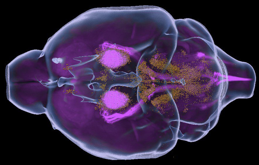
The supraspinal connectome is essential for normal behavior and homeostasis and consists of a wide range of sensory, motor, and autonomic projections from brain to spinal cord. Extensive work spanning a century has largely mapped the cell bodies of origin, yet their broad distribution and complex spatial relationships present significant challenges to the dissemination and application of this knowledge. Fields that study disruptions of supraspinal projections, for example spinal cord injury, have focused mostly on a handful of major populations that carry motor commands, with only limited consideration of dozens more that provide autonomic or crucial motor modulation.
The team felt that more comprehensive information was essential to understanding the functional consequences of different injuries and to better evaluate the efficacy of treatments. Using viral retrograde labeling, 3D imaging, and registration to standard neuro-anatomical atlases they have provided a platform to profile the entire supraspinal connectome by rapidly visualizing and quantifying tens of thousands of supraspinal neurons, each assigned to more than 60 identified regions and nuclei throughout the brains of adult mice. They then used this tool to compare the lumbar versus cervically-projecting connectomes, to profile brain-wide the sensitivity of supraspinal populations to graded spinal injuries, and to correlate locomotor recovery with connectome measurements. To freely share these insights in an intuitive manner, the team presented the information as an interactive web-based resource, which aims to spur progress by broadening understanding and analyses of essential but understudied supraspinal populations.
The research team presents a comprehensive and accessible approach to obtaining detailed information about the number and location of descending projection neurons throughout the mouse brain. This approach provides an effective tool to disseminate a comprehensive understanding of the supraspinal connectome, and a needed platform to rapidly achieve brain-wide profiling of disruption and restoration of brain-spinal cord connectivity after injury. Combined, these data establish an initial categorization and quantification of supraspinal brain regions in 3D space and create consistent experimental parameters for the detection of supraspinal neurons.
The paper and data presented in the website are from a collaborative project between Dr. Murray Blackmore’s Laboratory at Marquette University and Dr. Pantelis Tsoulfas’ Laboratory at The Miami Project to Cure Paralysis/University of Miami Miller School of Medicine. The work was funded by NIH/NINDS R01NS083983, The Miami Project and the Buoniconti fund.